Ultrasound Scan
An ultrasound scan, also known as sonography, is a non-invasive imaging technique that uses high-frequency sound waves to produce images of internal organs and tissues in real-time. This versatile diagnostic tool is widely used in various medical fields, including obstetrics, cardiology, and abdominal assessments. Ultrasound scans provide valuable information for diagnosing and monitoring a range of medical conditions without exposing patients to ionizing radiation.
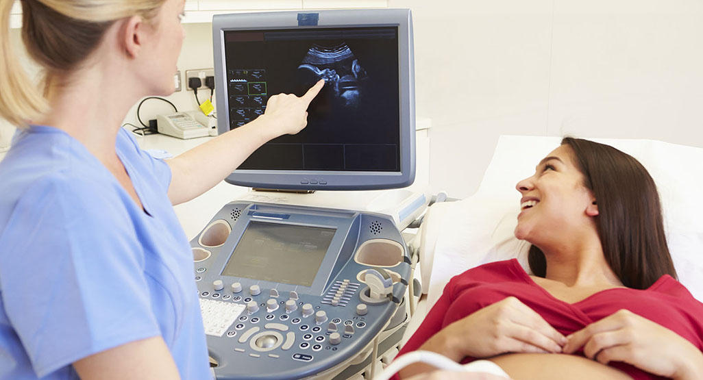
During an ultrasound scan, a small handheld device called a transducer is placed on the skin over the area of interest. The transducer emits sound waves that travel through the body and bounce back upon hitting different tissues. These echoes are then converted into visual images displayed on a monitor. The procedure is painless and typically takes between 15 to 30 minutes, depending on the area being examined. Patients may be asked to drink water before the scan to fill their bladder, which can enhance the visibility of certain organs.
Ultrasound scans can be classified into various types based on their applications. Obstetric ultrasound is commonly used to monitor fetal development during pregnancy, while abdominal ultrasound evaluates organs such as the liver, gallbladder, kidneys, and pancreas. Cardiac ultrasound, or echocardiography, assesses heart function and blood flow, while Doppler ultrasound measures blood flow in vessels, aiding in the diagnosis of vascular conditions.
One of the significant advantages of ultrasound scans is their safety profile. Unlike X-rays and CT scans, ultrasound does not use ionizing radiation, making it a safer option for pregnant women and children. Furthermore, ultrasound can be performed in a variety of settings, including outpatient clinics, hospitals, and at the bedside, increasing its accessibility for patients.
While ultrasound is an effective diagnostic tool, it does have limitations. Factors such as body habitus, the presence of gas in the intestines, or certain medical conditions can affect the quality of the images obtained. In some cases, additional imaging studies may be necessary for a comprehensive evaluation. However, advances in ultrasound technology, including 3D and 4D imaging, have significantly improved the clarity and detail of the images produced.
In conclusion, ultrasound scans are a crucial component of modern diagnostic medicine. Their non-invasive nature, safety, and versatility make them essential for evaluating a wide range of medical conditions. Regular use of ultrasound contributes to timely diagnosis and effective management of various health issues, enhancing patient care and outcomes.
- Non-Invasive: Ultrasound scans are painless and pose minimal risk to patients, making them suitable for all age groups.
- Real-Time Imaging: Provides immediate visualization of internal structures, facilitating quick diagnosis and treatment decisions.
- Diverse Applications: Used in various medical specialties, including obstetrics, cardiology, and emergency medicine.
- No Radiation Exposure: Unlike other imaging methods, ultrasound does not use ionizing radiation, ensuring safety for vulnerable populations.